Funke Lab Funke Lab

Overview

Our lab at the Human Technopole develops machine learning methods for the life sciences, with a focus on microscopy image analysis.

We are particularly interested in:

  1. Identification of Structures of Interest in Large Datasets

    In the field of connectomics, we develop methods to segment neurons, detect synapses, and to classify synapses in very large electron microscopy datasets.

    We also work on the segmentation and tracking of cells in live-cell imaging datasets.

  2. Explainable Machine Learning Methods

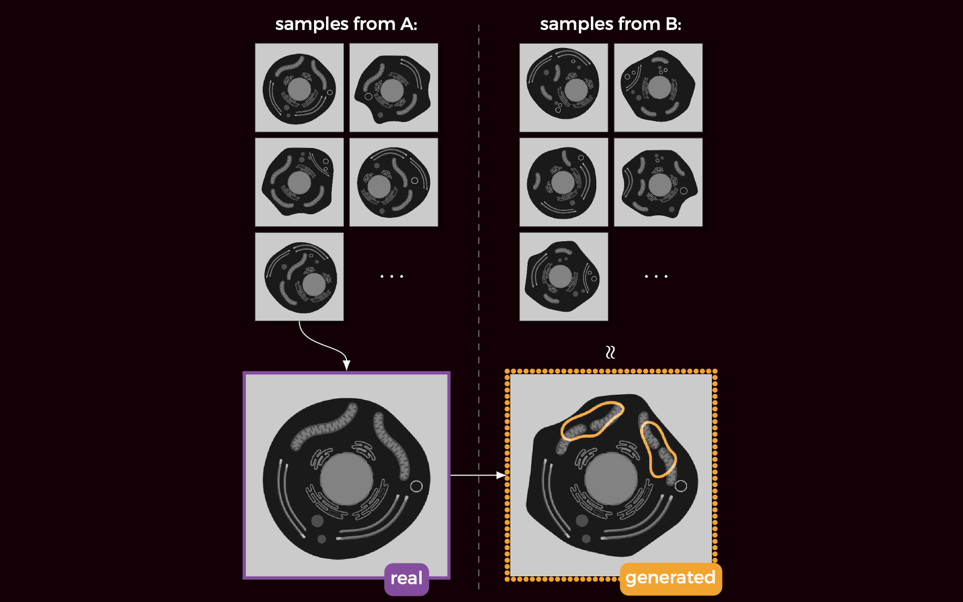
    We develop methods that use machine learning to identify and visualize subtle patterns in biological datasets. Those methods can reveal previously unknown phenotypical differences, e.g., in image data.

  3. Mechanistic Machine Learning

    To increase the utility and interpretability of machine learning methods, we design models that directly incorporate biophysical constraints and domain knowledge. So far, our models have been used to count fluorophores beyond the diffraction limit and to infer synaptic plasticity rules from behavioral measurements.

Selected publications

blinx

A Bayesian Model to Count the Number of Two-State Emitters in a Diffraction Limited Spot

Alexander Hillsley, Johannes Stein, Paul W. Tillberg, David L. Stern, Jan Funke

Nano Letters · 2025

DOI Paper Code

Abstract

We address the problem of inferring the number of independently blinking fluorescent light emitters, when only their combined intensity contributions can be observed. This problem occurs regularly in light microscopy of objects smaller than the diffraction limit, where one wishes to count the number of fluorescently labeled subunits. Our proposed solution directly models the photophysics of the system, as well as the blinking kinetics of the fluorescent emitters as a fully differentiable hidden Markov model, estimating a posterior distribution of the total number of emitters. We show that our model is more accurate and increases the range of countable subunits by a factor of 2 compared to current state-of-the-art methods. Furthermore, we demonstrate that our model can be used to investigate the effect of blinking kinetics on counting ability and therefore can inform optimal experimental conditions.

plastix

Model-Based Inference of Synaptic Plasticity Rules

Yash Mehta, Danil Tyulmankov, Adithya E. Rajagopalan, Glenn C. Turner, James E. Fitzgerald, Jan Funke

NeurIPS · 2024

Code Blog

Abstract

Inferring the synaptic plasticity rules that govern learning in the brain is a key challenge in neuroscience. We present a novel computational method to infer these rules from experimental data, applicable to both neural and behavioral data. Our approach approximates plasticity rules using a parameterized function, employing either truncated Taylor series for theoretical interpretability or multilayer perceptrons. These plasticity parameters are optimized via gradient descent over entire trajectories to align closely with observed neural activity or behavioral learning dynamics. This method can uncover complex rules that induce long nonlinear time dependencies, particularly involving factors like postsynaptic activity and current synaptic weights. We validate our approach through simulations, successfully recovering established rules such as Oja's, as well as more intricate plasticity rules with reward-modulated terms. We assess the robustness of our technique to noise and apply it to behavioral data from Drosophila in a probabilistic reward-learning experiment. Notably, our findings reveal an active forgetting component in reward learning in flies, improving predictive accuracy over previous models. This modeling framework offers a promising new avenue for elucidating the computational principles of synaptic plasticity and learning in the brain.

synister

Neurotransmitter Classification from Electron Microscopy Images at Synaptic Sites in Drosophila Melanogaster

Nils Eckstein, Alexander Shakeel Bates, Andrew Champion, Michelle Du, Yijie Yin, Philipp Schlegel, Alicia Kun-Yang Lu, Thomson Rymer, Samantha Finley-May, Tyler Paterson, Ruchi Parekh, Sven Dorkenwald, Arie Matsliah, Szi-Chieh Yu, Claire McKellar, Amy Sterling, Katharina Eichler, Marta Costa, Sebastian Seung, Mala Murthy, Volker Hartenstein, Gregory S. X. E. Jefferis, Jan Funke

Cell · 2024

DOI Paper Code

quac

Quantitative Attributions with Counterfactuals

Diane-Yayra Adjavon, Nils Eckstein, Alexander S. Bates, Gregory S. X. E. Jefferis, Jan Funke

bioRxiv · 2024

DOI Paper Code Website Blog

Abstract

We address the problem of explaining the decision process of deep neural network classifiers on images, which is of particular importance in biomedical datasets where class-relevant differences are not always obvious to a human observer. Our proposed solution, termed quantitative attribution with counterfactuals (QuAC), generates visual explanations that highlight class-relevant differences by attributing the classifier decision to changes of visual features in small parts of an image. To that end, we train a separate network to generate counterfactual images (i.e., to translate images between different classes). We then find the most important differences using novel discriminative attribution methods. Crucially, QuAC allows scoring of the attribution and thus provides a measure to quantify and compare the fidelity of a visual explanation. We demonstrate the suitability and limitations of QuAC on two datasets: (1) a synthetic dataset with known class differences, representing different levels of protein aggregation in cells and (2) an electron microscopy dataset of D. melanogaster synapses with different neurotransmitters, where QuAC reveals so far unknown visual differences. We further discuss how QuAC can be used to interrogate mispredictions to shed light on unexpected inter-class similarities and intra-class differences.

lsds

Local Shape Descriptors for Neuron Segmentation

Arlo Sheridan, Tri M. Nguyen, Diptodip Deb, Wei-Chung Allen Lee, Stephan Saalfeld, Srinivas C. Turaga, Uri Manor, Jan Funke

Nature Methods · 2023

DOI Paper Code Website

Abstract

We present an auxiliary learning task for the problem of neuron segmentation in electron microscopy volumes. The auxiliary task consists of the prediction of local shape descriptors (LSDs), which we combine with conventional voxel-wise direct neighbor affinities for neuron boundary detection. The shape descriptors capture local statistics about the neuron to be segmented, such as diameter, elongation, and direction. On a study comparing several existing methods across various specimen, imaging techniques, and resolutions, auxiliary learning of LSDs consistently increases segmentation accuracy of affinity-based methods over a range of metrics. Furthermore, the addition of LSDs promotes affinity-based segmentation methods to be on par with the current state of the art for neuron segmentation (flood-filling networks), while being two orders of magnitudes more efficient—a critical requirement for the processing of future petabyte-sized datasets.